Levels Of Support Pelvic Floor
The Pelvic Floor Northwest Wellness Group
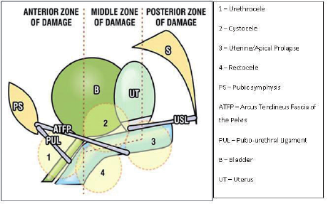
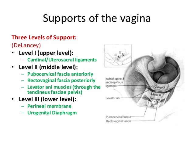
Three Levels Of Pelvic Support 5 Download Scientific Diagram

Picture Uterine Prolapse Pelvic Floor Exercises Medical Memes

Pelvic Organ Prolapse And Why Kegels Aren T Enough Pelvic Organ Prolapse Pelvic Floor Kegel

Your Pelvic Floor
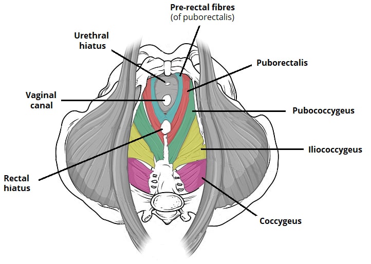
The Pelvic Floor Structure Function Muscles Teachmeanatomy

Huddleston ht dunnihoo dr huddleston pm 3rd meyers pc sr.
Levels of support pelvic floor.
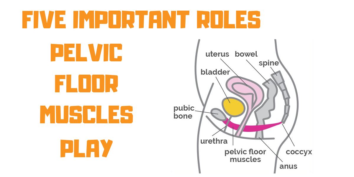
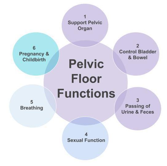
Pelvic Floor Muscles Five Important Roles Propel Physiotherapy

Lift Pelvic Support Series Femfusion Fitness In 2020 Pelvic Floor Pelvic Organ Prolapse Rehabilitation Exercises

Pin On Pelvic Floor

Pin On Incontinence Tips

Pelvic Organ Prolapse Pop Is Defined As A Relaxation Or Lack Of Support Via The Fascial And Ligam Pelvic Organ Prolapse Pelvic Floor Pelvic Floor Dysfunction

5 Ways To Reduce Your Risk Of Urogynecologic Health Issues Infographic Urinary Incontinence Infographic Health Incontinence
Pelvic Organ Prolapse

Kegel8 Blog 4 Simple Changes To Make Your Pelvic Floor Muscles Stronger Kegel8 Pelvic Floor Pelvic Floor Exercises Pelvic Floor Dysfunction

Kegel Exerciser Perifit Perifit In 2020 Kegel Exerciser Kegel Kegel Exercise

Free Resources The Tummy Team Online Core Rehabilitation Pelvic Floor Pelvic Floor Muscles Team Online

Three Damaging Pelvic Floor Rehab Myths Pelvic Floor Pelvic Organ Prolapse Pelvic Floor Dysfunction

Dealing With Stress Urinary Incontinence Don T Be Embarrassed Its Common And Treatable Urinary Incontinence Incontinence Dealing With Stress

Muscles At The Base Of The Pelvis Support Organs Of The Lower Abdomen In Women That Pelvi Pelvic Floor Dysfunction Pelvic Floor Therapy Pelvic Floor Prolapse

Pelvic Organ Prolapse And Why Kegels Aren T Enough Pelvic Floor Urinary Incontinence Body Confidence
Source : pinterest.com
